4D Ultrasound is a volumetric real-time ultrasound examination, allowing parents to observe the movements, facial expressions, and behavior of the baby. This method is safe, informative, and used for monitoring fetal development while creating an emotional connection before birth.
4D scanning provides a real-time 3D image of the baby, showing their facial expressions, movements, and position in the womb. The procedure is safe and helps evaluate the development of the fetus, offering parents their first visual contact with the baby.
What is 4D Ultrasound?
Unlike traditional 2D ultrasound, 4D scanning provides a dynamic three-dimensional image—like a video recording in real time. This allows the doctor to assess the baby’s anatomical structures in detail, while parents can emotionally engage in the process and cherish the moment forever.
The method is completely safe for both the mother and the baby and can be performed at various stages of pregnancy.
What can 4D scanning show?
- Facial features and emotional expressions
- Hand, foot, and head movements
- Fetal position
- Heartbeat frequency
- Development of organs
- Baby’s gender (if desired)
Advantages of 4D Ultrasound
- “Live” image of the baby in real-time
- Aesthetic and emotional experience for the parents
- Ability to detect abnormalities early
- Photos and videos—either for memory or family archives
- Comfortable conditions for the procedure
- Safe at any stage of pregnancy
When should 4D Ultrasound be performed?
The most optimal and visually informative period is between the 22nd and 30th weeks of pregnancy. At this stage, the baby is actively moving, with recognizable facial features and expressions, and the amniotic fluid level is ideal for clear visualization.
Preparation for the procedure
- No special preparation is required
- It is recommended to have a light snack an hour before the procedure to increase fetal activity
- Avoid applying greasy creams on the abdomen the day before the appointment
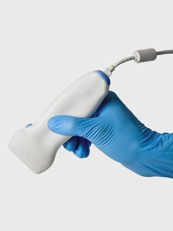
How the procedure is performed
The examination is conducted transabdominally using a sensor placed on the abdomen. A special gel is applied to the skin, and the doctor then displays the three-dimensional image of the baby in motion on the screen. The procedure lasts around 30–40 minutes and is peaceful and comfortable. If desired, parents can record photos and videos.