How the Examination Is Performed
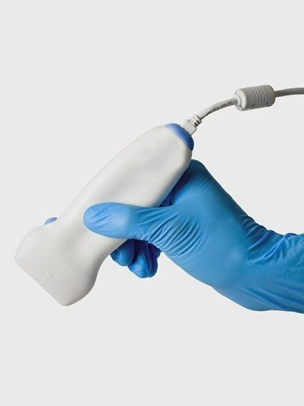
The procedure is performed in comfortable conditions and does not require special preparation. The patient lies on an examination table while a special gel is applied to the area being examined to improve ultrasound wave transmission.
During the procedure, the doctor moves the ultrasound probe along the skin and evaluates the structure of the vessels on the monitor. Ultrasound of veins and arteries allows visualization of vessel walls, measurement of vessel diameter, and detection of possible abnormalities such as atherosclerotic plaques or blood clots.
A Doppler mode is also used to evaluate the direction and speed of blood flow. This helps identify vessel narrowing, impaired circulation, and other blood flow disturbances.
Depending on medical indications, the examination may include ultrasound of neck vessels, limb vessels, or specific vascular segments. The procedure usually takes about 20–30 minutes.