Visual Diagnostics of Heart Function
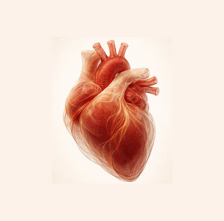
The heart is constantly in motion. During every heartbeat, valves open and close, blood flows through the chambers, and the myocardium performs complex coordinated work. To objectively evaluate these processes, ultrasound diagnostics are used.
Echocardiography is a modern diagnostic method that allows doctors to visualize the structure of the heart and assess its functional state in real time. Using ultrasound waves, the specialist can determine the size of the heart chambers, evaluate the condition of the valves, and assess the characteristics of blood flow.
This examination is often performed together with functional diagnostic methods such as an ECG. Electrocardiography shows the electrical activity of the myocardium, while heart ultrasound allows doctors to visually evaluate the anatomy and mechanical function of the heart muscle.
How the Examination Is Performed
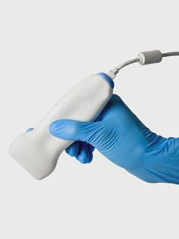
No special preparation is required before the procedure. The patient lies on an examination couch, and a special gel is applied to the chest area to improve ultrasound wave transmission.
During the examination, the doctor places the ultrasound probe on specific points of the chest and gradually evaluates different structures of the heart. Echocardiography allows detailed visualization of heart chambers, septa, valves, and major vessels. The monitor displays a dynamic image that shows the heart working in real time.
Blood flow inside the heart chambers is also evaluated. For this purpose, a Doppler mode is used to determine the direction and speed of blood flow.


")
")