Clinic «REFRESH»
Book a consultation
Book
area-icon
Dubai
English
menu_item_arrow
10:00 – 21:00 Sunday is a day off
Vascular Ultrasound Vascular Ultrasound

Vascular Ultrasound

A non-invasive ultrasound examination of arteries and veins that helps detect atherosclerosis, vascular narrowing, blood clots, and blood flow disturbances. Vascular ultrasound allows doctors to evaluate the condition of the vascular system and helps prevent the development of complications. Vascular ultrasound in Dubai is widely used for both preventive screening and diagnosis of vascular disorders.
Get consultation
Get consultation
Procedure Description

When It Is Important to Evaluate Vascular Health


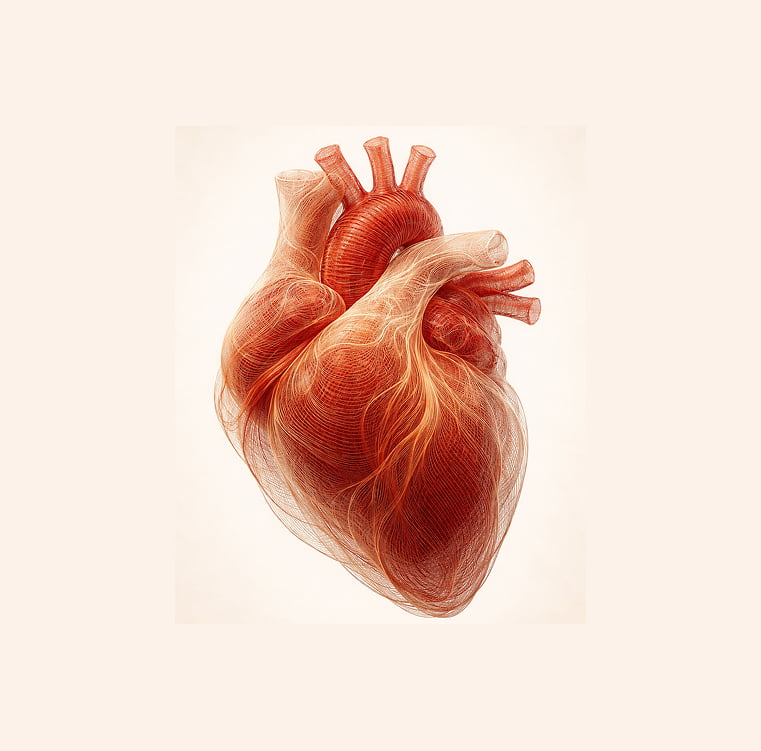
Blood vessels deliver oxygen and nutrients to all organs and tissues. Any structural changes or reduced vessel patency may gradually affect overall health, wellbeing, and physical activity levels.

Vascular ultrasound is a modern diagnostic method that allows doctors to evaluate arteries and veins without invasive procedures. Using ultrasound technology, specialists can assess vessel patency, detect changes in vessel walls, and analyze characteristics of blood flow.

The examination may be performed in the presence of various symptoms and is also used for preventive evaluation of the vascular system. For example, ultrasound of the neck vessels helps assess blood supply to the brain, while ultrasound of leg vessels can detect signs of varicose veins, thrombosis, or impaired venous circulation.

How the Examination Is Performed


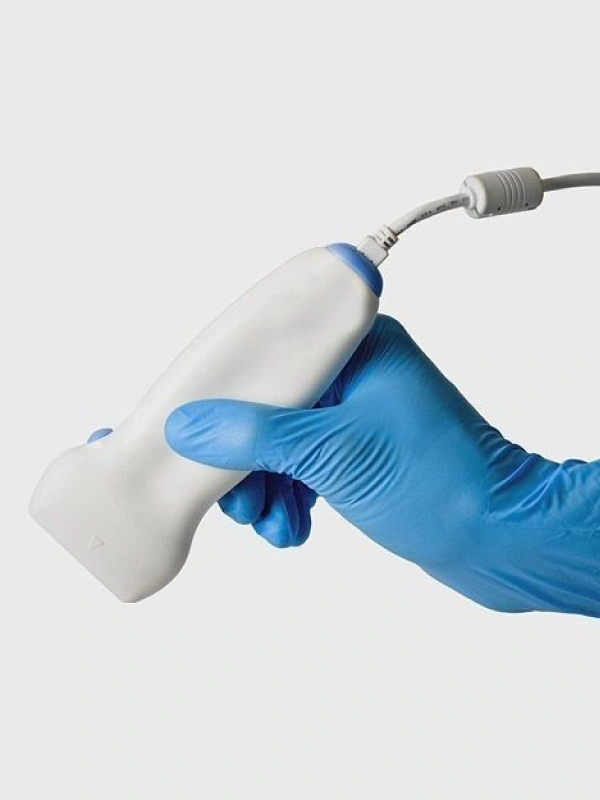
The procedure is performed in comfortable conditions and does not require special preparation. The patient lies on an examination table while a special gel is applied to the area being examined to improve ultrasound wave transmission.

During the procedure, the doctor moves the ultrasound probe along the skin and evaluates the structure of the vessels on the monitor. Ultrasound of veins and arteries allows visualization of vessel walls, measurement of vessel diameter, and detection of possible abnormalities such as atherosclerotic plaques or blood clots.

A Doppler mode is also used to evaluate the direction and speed of blood flow. This helps identify vessel narrowing, impaired circulation, and other blood flow disturbances.

Depending on medical indications, the examination may include ultrasound of neck vessels, limb vessels, or specific vascular segments. The procedure usually takes about 20–30 minutes.

What the Examination Can Detect


Vascular ultrasound provides detailed evaluation of both arterial and venous circulation. The method helps detect structural changes in vessel walls, identify atherosclerotic plaques, and analyze blood flow characteristics.

The examination plays an important role in early diagnosis of vascular diseases. For example, ultrasound of neck vessels can reveal changes in the carotid arteries that may affect blood supply to the brain, while ultrasound of leg vessels is commonly used to diagnose varicose veins and thrombosis.

Due to its high diagnostic value, vascular ultrasound is widely used for preventive screening and monitoring of vascular health in patients with chronic conditions.

Result
Evaluation of the structure of arteries and veins
Detection of atherosclerotic changes
Diagnosis of vascular narrowing
Identification of blood clots
Assessment of blood flow speed and direction
Evaluation of vessel wall condition
Indications
plus-icon
Pain or heaviness in the legs
plus-icon
Swelling of the lower limbs
plus-icon
Dizziness or ringing in the ears
plus-icon
Suspected atherosclerosis
plus-icon
Varicose vein disease
plus-icon
Preventive vascular health examination
Contraindications
minus-icon
The method is safe and non-invasive and therefore has no absolute contraindications. Limitations may include skin damage in the examination area or severe general medical conditions.