Visual Diagnostics of Digestive System Organs
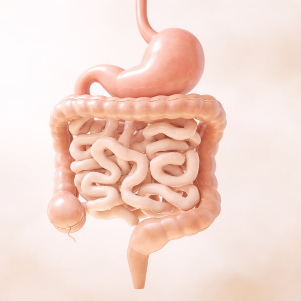
The organs of the abdominal cavity participate in complex processes of digestion, metabolism, and detoxification. Disturbances in their function may present with symptoms such as abdominal discomfort, heaviness after meals, or digestive changes.
Abdominal ultrasound is one of the primary diagnostic methods used to assess digestive system disorders. Using ultrasound waves, the doctor obtains real-time images of internal organs and evaluates their structure, size, and functional characteristics.
The examination helps identify changes in the liver, evaluate the condition of the gallbladder, pancreas, and spleen. Abdominal ultrasound can detect signs of inflammatory processes, bile flow disturbances, and other changes that may affect the gastrointestinal system.
How the Examination Is Performed
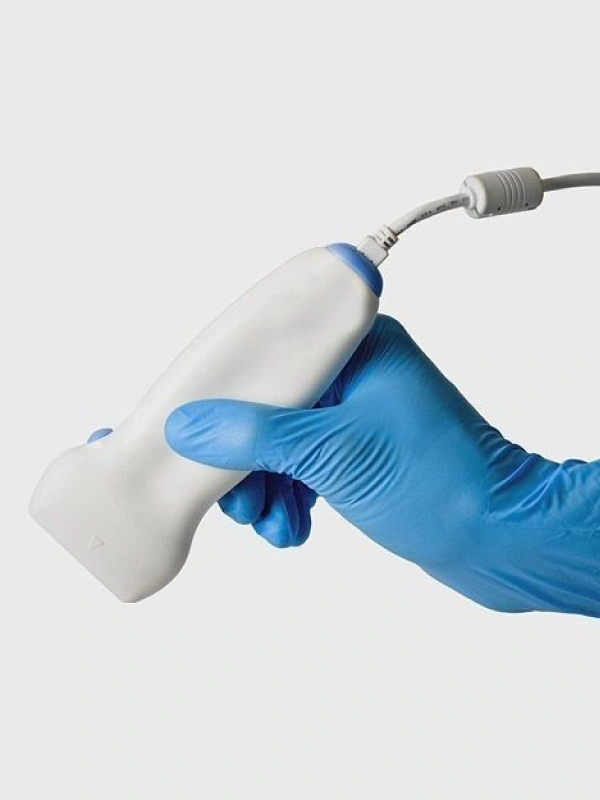
The procedure is performed in comfortable conditions and usually takes about 20–30 minutes. The patient lies on an examination table while a special gel is applied to the skin of the abdomen to improve ultrasound wave transmission.
During the procedure, the doctor moves a diagnostic probe across the abdominal surface and evaluates the condition of internal organs. Abdominal ultrasound allows detailed visualization of the liver, gallbladder, pancreas, and spleen.
During the examination, the specialist evaluates organ size, tissue structure, signs of inflammation, and the condition of the biliary system.
In some cases, preparation before abdominal ultrasound may be required. This may include following a specific diet before the examination to improve diagnostic accuracy and organ visualization.